Clinical protocol
PRF — platelet-rich fibrin
From blood draw to sticky bone: the complete PRF protocol applied to implant dentistry, trained on real cases in the Brazil residency.
The biomaterial that comes from the patient
PRF concentrates platelets, leukocytes and growth factors in a fibrin matrix obtained by centrifuging autologous blood — with no anticoagulants or additives. In implant dentistry it accelerates soft-tissue healing, improves particulate graft handling (sticky bone) and reduces post-operative morbidity. The clinical outcome depends on technical details: spin speed and time, time-to-use and membrane compression technique — exactly what we train in the residency.
Clinically reviewed in August 2026
L-PRF vs i-PRF: two forms, different uses
The centrifugation protocol defines the end product. Choosing the right form for each indication is the first step.
L-PRF (solid membranes)
- Low-speed centrifugation in a dry tube, without anticoagulant.
- Compressed into a membrane: covering grafts, sockets and soft-tissue defects.
- Sustained growth-factor release over 7 to 14 days.
i-PRF (liquid) and sticky bone
- Short, low-force spin: stays liquid for 10 to 15 minutes.
- Mixed with particulate graft it forms sticky bone, easy to mould and stabilise.
- Ideal for GBR, sinus lift and socket preservation.
The protocol in 5 phases
The exact sequence we perform in every residency surgery — from the patient's arm to the surgical field.
- 01
Blood draw
Venepuncture immediately before the critical surgical phase: less than 2 minutes between draw and centrifugation.
- 02
Centrifugation
Low-speed protocols (low-speed concept) for maximum cell concentration: balanced tubes, fixed rotor and calibrated times.
- 03
Membrane preparation
Separation of the fibrin clot and controlled compression in the PRF box to obtain homogeneous, resistant membranes.
- 04
Sticky bone
Hydrating particulate graft with liquid i-PRF: the resulting aggregate holds its shape, does not disperse and accelerates revascularisation.
- 05
Clinical application
Membranes over grafts and sockets, sticky bone in the defects and tension-free suturing: PRF only works with correct soft-tissue management.
Frequently asked questions
Does PRF replace bone graft?
No. PRF is not a filler material: it is a biological accelerator. In defects requiring volume it is combined with particulate graft (sticky bone) or barrier membranes.
What equipment is needed to make PRF?
A fixed-rotor centrifuge calibrated for low-speed protocols, specific additive-free tubes and a compression box for the membranes. A modest investment compared with the clinical benefit.
In which residency surgeries is PRF used?
In nearly all of them: GBR, sinus lift, socket preservation, immediate implants and All-on-4. Each participant prepares and applies their own PRF under supervision.
Are there contraindications to PRF?
Coagulation disorders, thrombocytopenia and unadjusted anticoagulant therapy are the main limitations. Pre-operative medical assessment is part of the residency protocol.
Who signs this protocol
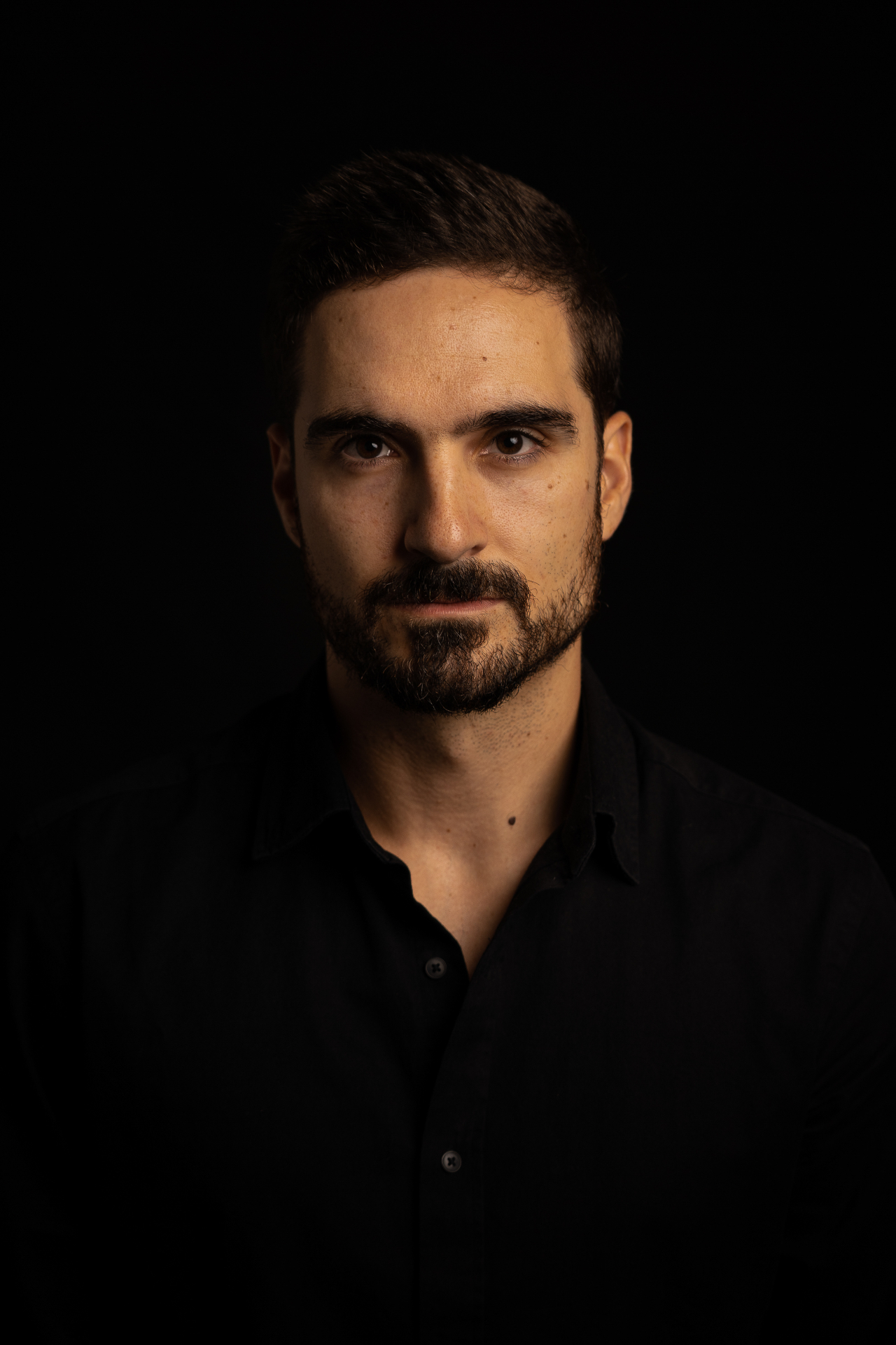
Dr. João Borges Nogueira
Oral surgeon practising in Paris, dedicated to advanced implantology, regeneration and international training of dentists.
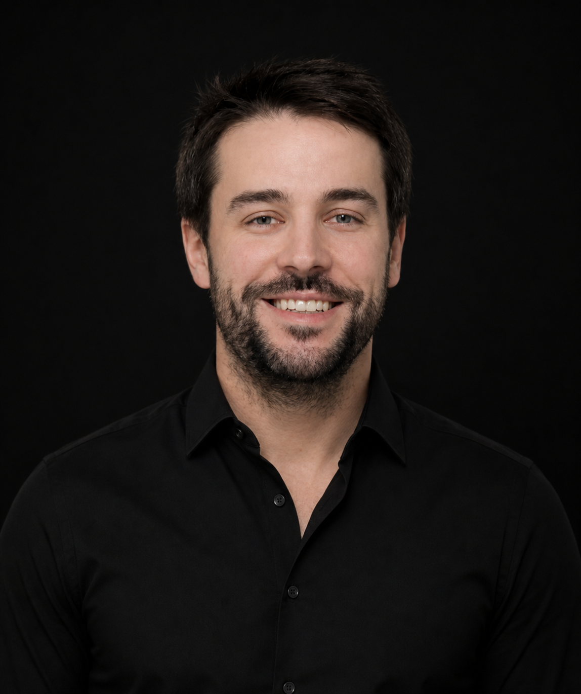
Dr. Gonçalo Castilho
Specialist in oral implantology practising in Portugal, focused on full-arch rehabilitations, digital planning and complex reconstruction cases.
Related procedures
These techniques combine within the same treatment plan — explore each protocol in detail.
All-on-4 — full-arch rehabilitation
Full-arch protocol on four implants with immediate loading.
Sinus lift
Lateral and transcrestal approaches to regain posterior bone height.
Guided bone regeneration (GBR)
Grafts and membranes to rebuild bone volume before or during implant placement.
Immediate post-extraction implants
Placement in the same surgical session, managing the gap and emergence profile.
Master PRF in practice, not just in theory
In the intensive Brazil residency you will prepare and apply PRF in real regenerative surgeries, alongside the faculty.